|
|
Barton Fracture
General Considerations
- Intra-articular fracture of the distal radius with subluxation/dislocation of the wrist
- The lunate maintains its relationship with the fracture fragment
- May involve the volar aspect of the radius (sometimes called “reverse Barton fracture”) or the dorsal aspect
- Volar type is more common
- Most common fracture-dislocation of the wrist
Clinical Findings
- Dorsal Barton’s fracture usually results from fall on an out-stretched hand
- Volar-type is caused by the same mechanism as a Smith fracture
- Result is deformity at wrist joint
- Pain, tenderness, swelling and decreased range of motion
Imaging Findings
- Conventional radiographs are the study of first choice
- Fracture is wedge-shaped and extends obliquely on the lateral view into the radiocarpal joint
- Carpal bones and hand maintain their relationship with the fracture fragment, separating from the remainder of the radius
- Frequently associated with ulnar styloid fractures
- CT if further definition is required
Differential Diagnosis
- Colles fracture-extra-articular fracture of the distal radius with dorsal angulation
- Smith fracture-a fracture of the distal radius with palmar angulation
- May have intra-articular component, but dislocation is not a feature as in Barton fracture
Treatment
- Most require operative treatment
Prognosis
- May re-dislocate
- Malunion

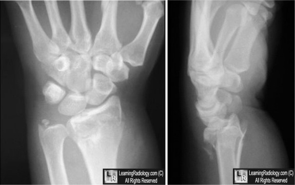
Barton's Fracture-Dislocation of the Wrist. The frontal radiograph shows a
fracture of the
distal radius (red arrow) and ulnar styloid (white arrow). The lateral radiograph demonstrates
a dorsal, wedge-shaped, intra-articular fracture of the
distal radius (yellow arrow),
with which the carpal bones (green arrow) have dislocated posteriorly.
For these same photos without the arrows, click here
For more information, click on the link if you see this icon 
Barton fractures. Wheeless' Textbook of Orthopaedics
|
|
|
